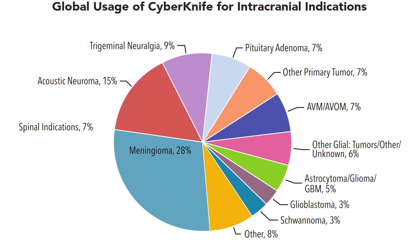
AVMs
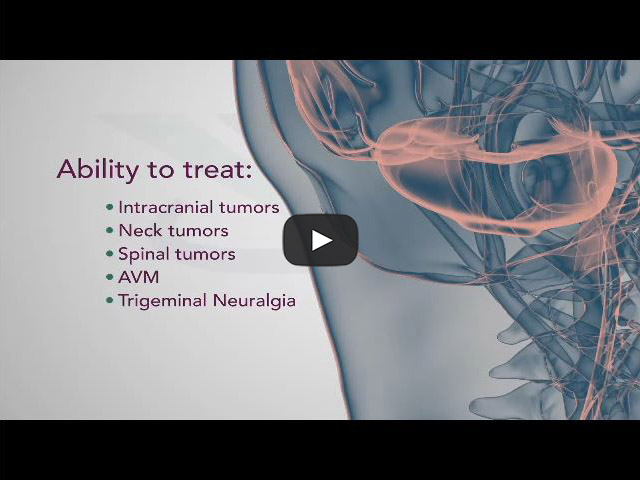
CyberKnife explained by Neurosurgeons
CyberKnife vs. Gamma Knife
The CyberKnife System and Gamma Knife are two of the treatment options for AVM. Find out more about the differences between CyberKnife and Gamma Knife here.
What are arteriovenous malformations? (AVM)
Arteriovenous malformations (AVMs) are abnormal, snarled tangles of blood vessels that cause multiple irregular connections between the arteries and veins. While they are most common in the spinal cord and the brain, but can develop elsewhere in the body. It is unclear why AVMs form. Most often AVMs are congenital, but they can appear sporadically. In some cases the AVM may be inherited, but it is more likely that other inherited conditions increase the risk of having an AVM.
Normally, arteries carry oxygen-rich blood away from the heart to the body’s cells, organs, and tissues; veins return blood with less oxygen to the lungs and heart. But in an AVM, the absence of capillaries—a network of small blood vessels that connect arteries to veins and deliver oxygen to cells—creates a shortcut for blood to pass directly from arteries to veins and bypass tissue, which can lead to tissue damage and the death of nerve cells and other cells. Over time, some AVMs get progressively larger as the amount of blood flow increases.
What are the symptoms?
AVM symptoms can vary greatly in severity; in some people the severity of symptoms becomes debilitating or even life threatening.
Seizures and headaches that may be severe are the most generalized symptoms of AVMs, but no particular type of seizure or headache pattern has been identified. Seizures can be focal (meaning they involve a small part of the brain) or generalized (widespread), involving convulsions, a loss of control over movement or a change in a person’s level of consciousness. Headaches can vary greatly in frequency, duration, and intensity, sometimes becoming as severe as migraines. Pain may be on either one side of the head or on both sides. Sometimes a headache consistently affecting one side of the head may be closely linked to the site of an AVM. Most often, the location of the pain is not specific to the malformation and may encompass most of the head.
AVMs also can cause a wide range of more specific neurological symptoms that vary from person to person, depending primarily upon the location of the AVM:
- muscle weakness or paralysis in one part of the body
- a loss of coordination (ataxia) that can lead to such problems as gait disturbances
- difficulties carrying out tasks that require planning
- back pain or weakness in the lower extremities caused by a spinal AVM
- dizziness
- visual problems such as a loss of part of the visual field, inability to control eye movement, or swelling of a part of the optic nerve
- difficulty speaking or understanding language (aphasia)
- abnormal sensations such as numbness, tingling, or spontaneous pain
- memory deficits
- confusion, hallucinations or dementia.
AVMs also may cause subtle learning or behavioral disorders in some people during their childhood or adolescence, long before more obvious symptoms become evident.
How do AVMs damage the brain and spinal cord?
AVMs damage the brain or spinal cord through three basic mechanisms:
- Reducing the amount of oxygen reaching neurological tissues
- Causing bleeding (hemorrhage) into surrounding tissues
- Compressing or displacing parts of the brain or spinal cord
Where do neurological AVMs tend to form?
AVMs can form virtually anywhere in the brain or spinal cord—wherever arteries and veins exist. Some are formed from blood vessels located in the outermost (dura mater) and innermost (pia mater) of the three membranes surrounding the brain and spinal cord. (The third membrane, called the arachnoid, lacks blood vessels.) Spinal AVMs frequently cause attacks of sudden, severe back pain, often concentrated at the roots of nerve fibers where they exit the vertebrae, with pain that is similar to that caused by a slipped disk. These lesions also can cause sensory disturbances, muscle weakness or paralysis in the parts of the body served by the spinal cord or the damaged nerve fibers. A spinal cord AVM can lead to degeneration of the nerve fibers within the spinal cord below the level of the lesion, causing widespread paralysis in parts of the body controlled by those nerve fibers.
AVMs on the surface of the cerebral hemispheres—the uppermost portions of the brain—exert pressure on the cerebral cortex, the brain’s “gray matter.” Depending on their location, these AVMs may damage portions of the cerebral cortex involved with thinking, speaking, understanding language, hearing, taste, touch, or initiating and controlling voluntary movements. AVMs located on the frontal lobe close to the optic nerve or on the occipital lobe (the rear portion of the cerebrum where images are processed) may cause a variety of visual problems.
AVMs also can form from blood vessels located deep inside the interior of the cerebrum (the main portion of the brain). These AVMs may compromise the functions of three vital structures: the thalamus, which transmits nerve signals between the spinal cord and upper regions of the brain; the basal ganglia surrounding the thalamus, which coordinate complex movements and plays a role in learning and memory; and the hippocampus, which plays a major role in memory.
AVMs can affect other parts of the brain besides the cerebrum. The hindbrain is formed from two major structures: the cerebellum, which is nestled under the rear portion of the cerebrum, and the brain stem, which serves as the bridge linking the upper portions of the brain with the spinal cord. These structures control finely coordinated movements, maintain balance, and regulate some functions of internal organs, including those of the heart and lungs. AVM damage to these parts of the hindbrain can result in dizziness, giddiness, vomiting, a loss of the ability to coordinate complex movements such as walking, or uncontrollable muscle tremors.
What are the health consequences of AVMs?
The greatest potential danger posed by AVMs is hemorrhage. Most episodes of bleeding are undetected at the time they occur because they are not severe enough to cause significant neurological damage. But massive, even fatal, bleeding episodes do occur. Whenever an AVM is detected, the individual should be carefully and consistently monitored for any signs of instability that may indicate an increased risk of hemorrhage.
A few physical characteristics appear to indicate a greater-than-usual likelihood of clinically significant hemorrhage:
- Smaller AVMs have a greater likelihood of bleeding than do larger ones.
- Impaired drainage by unusually narrow or deeply situated veins increases the chances of hemorrhage.
- Pregnancy appears to increase the likelihood of clinically significant hemorrhage, mainly because of increases in blood pressure and blood volume.
- AVMs that have hemorrhaged once are about nine times more likely to bleed again during the first year after the initial hemorrhage than are lesions that have never bled.
The damaging effects of a hemorrhage are related to lesion location. Bleeding from AVMs located deep inside the interior tissues, or parenchyma, of the brain typically causes more severe neurological damage than does hemorrhage by lesions that have formed in the dural or pial membranes or on the surface of the brain or spinal cord. Therefore, location is an important factor to consider when weighing the relative risks surgery to treat AVMs.
How are AVMs and other vascular lesions treated?
There are several options for treating AVMs. Although medication can often lessen general symptoms, such as headache, back pain, and seizures, the definitive treatment for AVMs is either surgery or focused radiation therapy.
Because so many variables are involved in treating AVMs, doctors must assess the danger posed to individuals largely on a case-by-case basis. A hemorrhage from an untreated AVM can cause serious neurological deficits or death, leading many clinicians to recommend surgical. However, surgery on any part of the central nervous system carries risk of serious complications or death. There is no easy formula that can allow physicians and individuals to reach a decision on the best course of therapy.
An AVM grading system developed in the mid-1980s can help health care professionals estimate the risk of surgery based on the size of the AVM, location in the brain and surrounding tissue involvement, and any leakage.
Treatment options include:
- Conventional surgery – Surgery entails entering the brain or spinal cord and removing the central portion of the AVM, including the fistula. This surgery is most appropriate when an AVM is located in a superficial portion of the brain or spinal cord and is relatively small in size. AVMs located deep inside the brain generally cannot be approached through conventional surgical techniques because there is too great a possibility that functionally important brain tissue will be damaged or destroyed.
- Endovascular embolization – For this treatment, a surgeon guides a catheter though the arterial network until the tip reaches the site of the AVM, then injects a fast-drying glue-like substance, fibered titanium coils or a tiny balloon) that will travel through blood vessels and create an artificial blood clot in the center of an AVM. Since embolization usually does not permanently obliterate the AVM, it is usually used as an adjunct to surgery or to radiosurgery to reduce the blood flow through the AVM and make the surgery safer.
- Radiosurgery – A less invasive therapeutic approach often used to treat small AVMs that haven’t ruptured is radiosurgery, in which a beam of highly focused radiation is aimed directly on the AVM and damages the walls of the blood vessels making up the lesion. Over the course of the next several months, the irradiated vessels gradually degenerate and eventually close, leading to the resolution of the AVM.
What causes vascular lesions?
The cause of vascular anomalies of the central nervous system is not yet well understood. Scientists believe the anomalies most often result from mistakes that occur during embryonic or fetal development, including genetic mutations. A few types of vascular malformations are known to be hereditary. Some lesions may also occur later in life as a result of injury to the central nervous system.
How are AVMs and other vascular lesions detected?
One of the more distinctive signs clinicians use to diagnose an AVM is an auditory phenomenon called a bruit—a rhythmic, whooshing sound caused by excessively rapid blood flow through the arteries and veins of an AVM. The sound is similar to a torrent of water rushing through a narrow pipe. When audible to individuals, the bruit may compromise hearing, disturb sleep, or cause significant psychological distress.
Various imaging technologies can be used to uncover the presence of AVMs:
- Cerebral angiography, also called cerebral arteriography, provides the most accurate pictures of blood vessel structure in brain AVMs. A special water-soluble dye, called a contrast agent, is injected into an artery and highlights the structure of blood vessels so that it can be seen on X-rays.
- CT or CAT scans (computed axial tomography) use X-rays to create an image of the head, brain or spinal cord and are especially useful in revealing the presence of hemorrhage.
- MRI (magnetic resonance imaging) uses magnetic fields and radio waves to create detailed images that can show subtle changes in neurological tissues.
- MRA (magnetic resonance angiography) can record the pattern and velocity of blood flow through vascular lesions, as well as the flow of cerebrospinal fluid throughout the brain and spinal cord.
- Transcranial Doppler ultrasound can diagnose medium-size to large AVMS and also detect the presence and extent of hemorrhage. It evaluates blood flow through the brain by directing high-frequency sound waves through the skull at particular arteries. The resulting sound wave signals that bounce back from blood cells are interpreted by a computer to make an image of the velocity of blood flow.
Patient Education

CyberKnife - Brain Cancer

TrueBeam STx - Brain, Head & Neck Cancer
CyberKnife explained by Neurosurgeons
Truebeam STx - Brain Tumors
Patient Testimonials
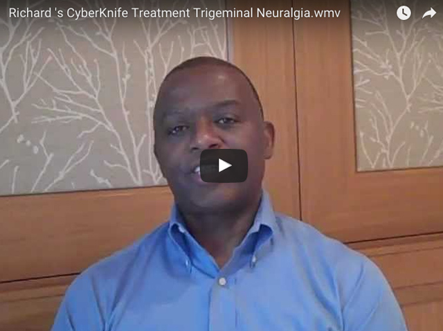
Richard 's CyberKnife Treatment Trigeminal Neuralgia

CyberKnife Treatment Acoustic Neuroma (AN)

Rachael's CyberKnife Treatment Chordoma Spinal Tumor
CyberKnife explained by Neurosurgeons
CyberKnife vs. Gamma Knife
The CyberKnife System and Gamma Knife are two of the treatment options for AVM. Find out more about the differences between CyberKnife and Gamma Knife here.

What are arteriovenous malformations? (AVM)
Arteriovenous malformations (AVMs) are abnormal, snarled tangles of blood vessels that cause multiple irregular connections between the arteries and veins. While they are most common in the spinal cord and the brain, but can develop elsewhere in the body. It is unclear why AVMs form. Most often AVMs are congenital, but they can appear sporadically. In some cases the AVM may be inherited, but it is more likely that other inherited conditions increase the risk of having an AVM.
Normally, arteries carry oxygen-rich blood away from the heart to the body’s cells, organs, and tissues; veins return blood with less oxygen to the lungs and heart. But in an AVM, the absence of capillaries—a network of small blood vessels that connect arteries to veins and deliver oxygen to cells—creates a shortcut for blood to pass directly from arteries to veins and bypass tissue, which can lead to tissue damage and the death of nerve cells and other cells. Over time, some AVMs get progressively larger as the amount of blood flow increases.
CyberKnife explained by Neurosurgeons
What are the symptoms?
AVM symptoms can vary greatly in severity; in some people the severity of symptoms becomes debilitating or even life threatening.
Seizures and headaches that may be severe are the most generalized symptoms of AVMs, but no particular type of seizure or headache pattern has been identified. Seizures can be focal (meaning they involve a small part of the brain) or generalized (widespread), involving convulsions, a loss of control over movement or a change in a person’s level of consciousness. Headaches can vary greatly in frequency, duration, and intensity, sometimes becoming as severe as migraines. Pain may be on either one side of the head or on both sides. Sometimes a headache consistently affecting one side of the head may be closely linked to the site of an AVM. Most often, the location of the pain is not specific to the malformation and may encompass most of the head.
AVMs also can cause a wide range of more specific neurological symptoms that vary from person to person, depending primarily upon the location of the AVM:
- muscle weakness or paralysis in one part of the body
- a loss of coordination (ataxia) that can lead to such problems as gait disturbances
- difficulties carrying out tasks that require planning
- back pain or weakness in the lower extremities caused by a spinal AVM
- dizziness
- visual problems such as a loss of part of the visual field, inability to control eye movement, or swelling of a part of the optic nerve
- difficulty speaking or understanding language (aphasia)
- abnormal sensations such as numbness, tingling, or spontaneous pain
- memory deficits
- confusion, hallucinations or dementia.
AVMs also may cause subtle learning or behavioral disorders in some people during their childhood or adolescence, long before more obvious symptoms become evident.
How do AVMs damage the brain and spinal cord?
AVMs damage the brain or spinal cord through three basic mechanisms:
- Reducing the amount of oxygen reaching neurological tissues
- Causing bleeding (hemorrhage) into surrounding tissues
- Compressing or displacing parts of the brain or spinal cord
Where do neurological AVMs tend to form?
AVMs can form virtually anywhere in the brain or spinal cord—wherever arteries and veins exist. Some are formed from blood vessels located in the outermost (dura mater) and innermost (pia mater) of the three membranes surrounding the brain and spinal cord. (The third membrane, called the arachnoid, lacks blood vessels.) Spinal AVMs frequently cause attacks of sudden, severe back pain, often concentrated at the roots of nerve fibers where they exit the vertebrae, with pain that is similar to that caused by a slipped disk. These lesions also can cause sensory disturbances, muscle weakness or paralysis in the parts of the body served by the spinal cord or the damaged nerve fibers. A spinal cord AVM can lead to degeneration of the nerve fibers within the spinal cord below the level of the lesion, causing widespread paralysis in parts of the body controlled by those nerve fibers.
AVMs on the surface of the cerebral hemispheres—the uppermost portions of the brain—exert pressure on the cerebral cortex, the brain’s “gray matter.” Depending on their location, these AVMs may damage portions of the cerebral cortex involved with thinking, speaking, understanding language, hearing, taste, touch, or initiating and controlling voluntary movements. AVMs located on the frontal lobe close to the optic nerve or on the occipital lobe (the rear portion of the cerebrum where images are processed) may cause a variety of visual problems.
AVMs also can form from blood vessels located deep inside the interior of the cerebrum (the main portion of the brain). These AVMs may compromise the functions of three vital structures: the thalamus, which transmits nerve signals between the spinal cord and upper regions of the brain; the basal ganglia surrounding the thalamus, which coordinate complex movements and plays a role in learning and memory; and the hippocampus, which plays a major role in memory.
AVMs can affect other parts of the brain besides the cerebrum. The hindbrain is formed from two major structures: the cerebellum, which is nestled under the rear portion of the cerebrum, and the brain stem, which serves as the bridge linking the upper portions of the brain with the spinal cord. These structures control finely coordinated movements, maintain balance, and regulate some functions of internal organs, including those of the heart and lungs. AVM damage to these parts of the hindbrain can result in dizziness, giddiness, vomiting, a loss of the ability to coordinate complex movements such as walking, or uncontrollable muscle tremors.
What are the health consequences of AVMs?
The greatest potential danger posed by AVMs is hemorrhage. Most episodes of bleeding are undetected at the time they occur because they are not severe enough to cause significant neurological damage. But massive, even fatal, bleeding episodes do occur. Whenever an AVM is detected, the individual should be carefully and consistently monitored for any signs of instability that may indicate an increased risk of hemorrhage.
A few physical characteristics appear to indicate a greater-than-usual likelihood of clinically significant hemorrhage:
- Smaller AVMs have a greater likelihood of bleeding than do larger ones.
- Impaired drainage by unusually narrow or deeply situated veins increases the chances of hemorrhage.
- Pregnancy appears to increase the likelihood of clinically significant hemorrhage, mainly because of increases in blood pressure and blood volume.
- AVMs that have hemorrhaged once are about nine times more likely to bleed again during the first year after the initial hemorrhage than are lesions that have never bled.
The damaging effects of a hemorrhage are related to lesion location. Bleeding from AVMs located deep inside the interior tissues, or parenchyma, of the brain typically causes more severe neurological damage than does hemorrhage by lesions that have formed in the dural or pial membranes or on the surface of the brain or spinal cord. Therefore, location is an important factor to consider when weighing the relative risks surgery to treat AVMs.
How are AVMs and other vascular lesions treated?
There are several options for treating AVMs. Although medication can often lessen general symptoms, such as headache, back pain, and seizures, the definitive treatment for AVMs is either surgery or focused radiation therapy.
Because so many variables are involved in treating AVMs, doctors must assess the danger posed to individuals largely on a case-by-case basis. A hemorrhage from an untreated AVM can cause serious neurological deficits or death, leading many clinicians to recommend surgical. However, surgery on any part of the central nervous system carries risk of serious complications or death. There is no easy formula that can allow physicians and individuals to reach a decision on the best course of therapy.
An AVM grading system developed in the mid-1980s can help health care professionals estimate the risk of surgery based on the size of the AVM, location in the brain and surrounding tissue involvement, and any leakage.
Treatment options include:
- Conventional surgery – Surgery entails entering the brain or spinal cord and removing the central portion of the AVM, including the fistula. This surgery is most appropriate when an AVM is located in a superficial portion of the brain or spinal cord and is relatively small in size. AVMs located deep inside the brain generally cannot be approached through conventional surgical techniques because there is too great a possibility that functionally important brain tissue will be damaged or destroyed.
- Endovascular embolization – For this treatment, a surgeon guides a catheter though the arterial network until the tip reaches the site of the AVM, then injects a fast-drying glue-like substance, fibered titanium coils or a tiny balloon) that will travel through blood vessels and create an artificial blood clot in the center of an AVM. Since embolization usually does not permanently obliterate the AVM, it is usually used as an adjunct to surgery or to radiosurgery to reduce the blood flow through the AVM and make the surgery safer.
- Radiosurgery – A less invasive therapeutic approach often used to treat small AVMs that haven’t ruptured is radiosurgery, in which a beam of highly focused radiation is aimed directly on the AVM and damages the walls of the blood vessels making up the lesion. Over the course of the next several months, the irradiated vessels gradually degenerate and eventually close, leading to the resolution of the AVM.
What causes vascular lesions?
The cause of vascular anomalies of the central nervous system is not yet well understood. Scientists believe the anomalies most often result from mistakes that occur during embryonic or fetal development, including genetic mutations. A few types of vascular malformations are known to be hereditary. Some lesions may also occur later in life as a result of injury to the central nervous system.
How are AVMs and other vascular lesions detected?
One of the more distinctive signs clinicians use to diagnose an AVM is an auditory phenomenon called a bruit—a rhythmic, whooshing sound caused by excessively rapid blood flow through the arteries and veins of an AVM. The sound is similar to a torrent of water rushing through a narrow pipe. When audible to individuals, the bruit may compromise hearing, disturb sleep, or cause significant psychological distress.
Various imaging technologies can be used to uncover the presence of AVMs:
- Cerebral angiography, also called cerebral arteriography, provides the most accurate pictures of blood vessel structure in brain AVMs. A special water-soluble dye, called a contrast agent, is injected into an artery and highlights the structure of blood vessels so that it can be seen on X-rays.
- CT or CAT scans (computed axial tomography) use X-rays to create an image of the head, brain or spinal cord and are especially useful in revealing the presence of hemorrhage.
- MRI (magnetic resonance imaging) uses magnetic fields and radio waves to create detailed images that can show subtle changes in neurological tissues.
- MRA (magnetic resonance angiography) can record the pattern and velocity of blood flow through vascular lesions, as well as the flow of cerebrospinal fluid throughout the brain and spinal cord.
- Transcranial Doppler ultrasound can diagnose medium-size to large AVMS and also detect the presence and extent of hemorrhage. It evaluates blood flow through the brain by directing high-frequency sound waves through the skull at particular arteries. The resulting sound wave signals that bounce back from blood cells are interpreted by a computer to make an image of the velocity of blood flow.